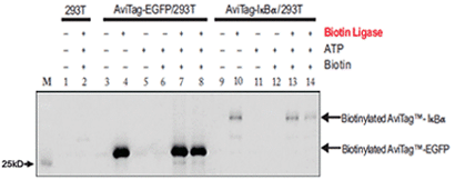
Example of in vitro biotinylation of AviTag recombinant proteins
Figure1. Extracts from 293T cells transfected with the AviTag eGFP or AviTag-IkBαexpression plasmid and untransfected controls were mixed with purified biotin ligase, biotin (50M) and ATP (10mM) as indicated in the figure. After incubation at 30.C (lanes 1-7, 9-13) or 4.C (lanes 8, 14) for 30 minutes, the mixtures were resolved by SDS-PAGE. Biotinylated proteins were visualized on a western blot with streptavidin-HRP conjugate and chemiluminescent substrate (Pierce).
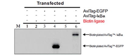
Example of in vivo biotinylation of AviTag recombinant proteins
Figure2. AviTag-eGFP or AviTag- IkBα expression plasmids were transfected into 293T cells alone or with co-transfected biotin ligase. Lysates were prepared 13 hours. Extracts from after transfection and resolved on a 12% SDS-PAGE gel. Biotinylated proteins were visualized on a western blot with streptavidin-HRP conjugate and chemiluminescent substrate (Pierce).
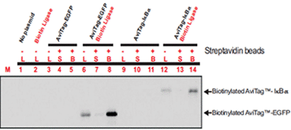
Example of precipitation of biotinylated AviTag recombinant proteins with streptavidin beads
Figure3. 293T cells were transfected with different combinations of AviTag expression plasmids and biotin ligase, as indicated in the figure. Lysates were prepared 24 hours after transfection and incubated with streptavidin beads for 8 hours. Suspensions were centrifuged and pelleted beads were washed 3 times. Cell lysates (L), supernatants (S) and pelleted biotinylated protein bound to streptavidin beads (B) were resolved by SDS-PAGE. Biotinylated proteins were visualized on a western blot with streptavidin-HRP conjugate and chemiluminescent substrate (Pierce).